noun, a person or thing that is mysterious or difficult to understand.
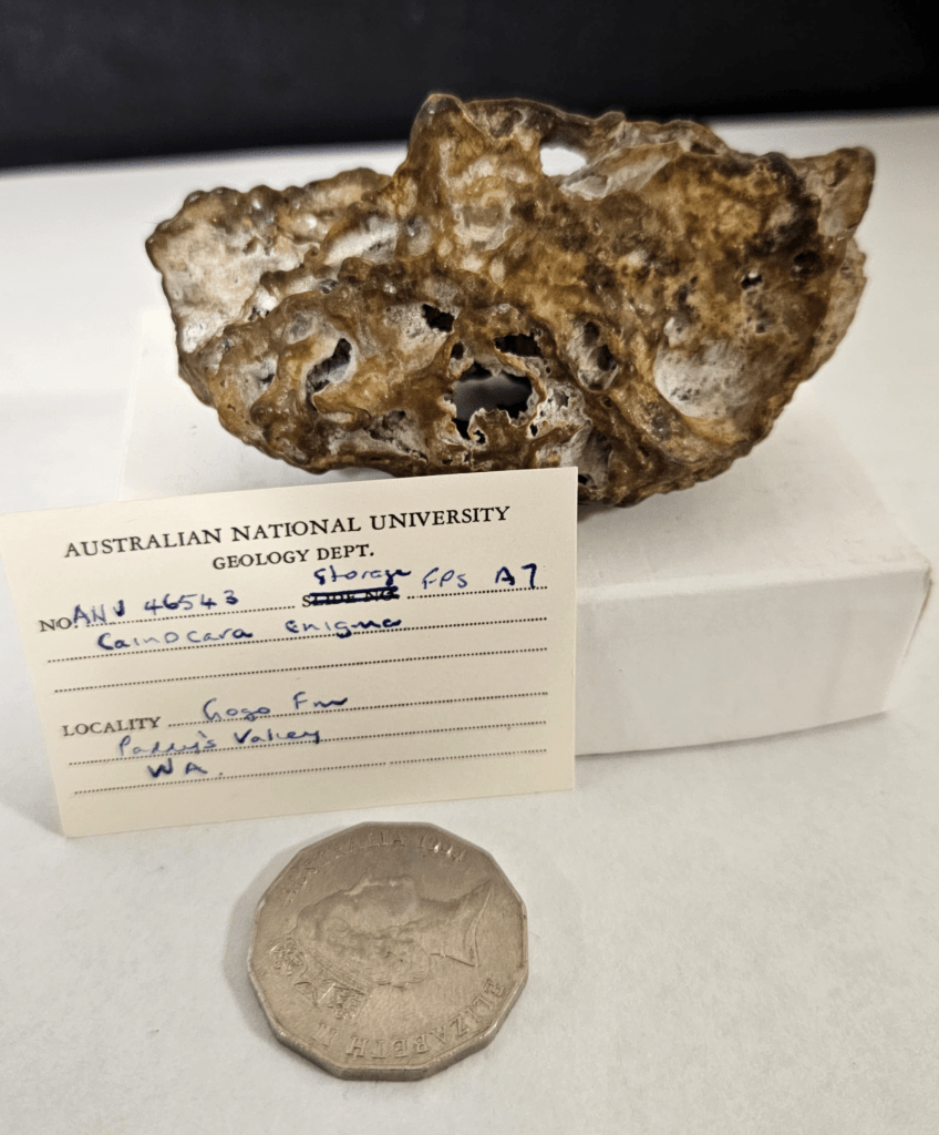
Since 2011, there has persisted a mysterious specimen known from the Late Devonian Gogo Formation in Australia. Dubbed Cainocara enigma by authors Ken Campbell and Dick Barwick, who first studied it, they published a work entitled “A new unusual Osteichthyan fish from the Gogo Formation, Western Australia“, but considered this single specimen so strange that they could not identify it any further than some kind of bony fish…
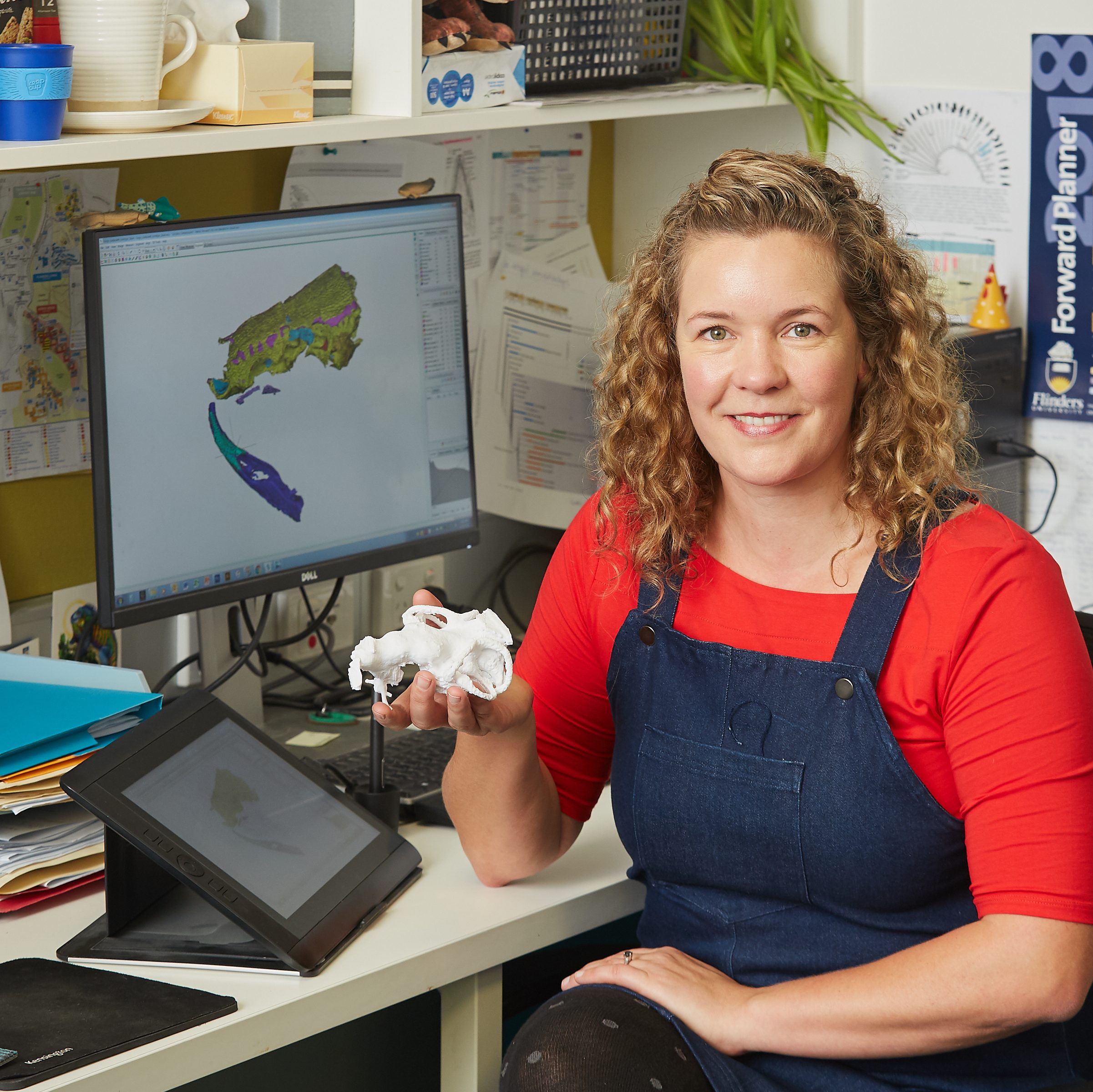
Fast forward to 2024, and enter my dedicated and hard-working 3rd year research student, Hannah Thiele, who used CT scans of this, well, BLOB of a fossil (there really is no other way to describe it!) and specialised 3D segmenting software to solve the mystery of just what the enigma really was!
A photo of the enigma from Gogo, housed in the Australian National University fossil collection.
By reconstructing the internal space of this mysterious fossil, Hannah was able to identify a cranial endocast, the space inside the skull that usually houses the brain. In doing so, we could identify features that helped us to orient the specimen correctly and interpret it accurately. This meant we could solve the mystery once and for all!
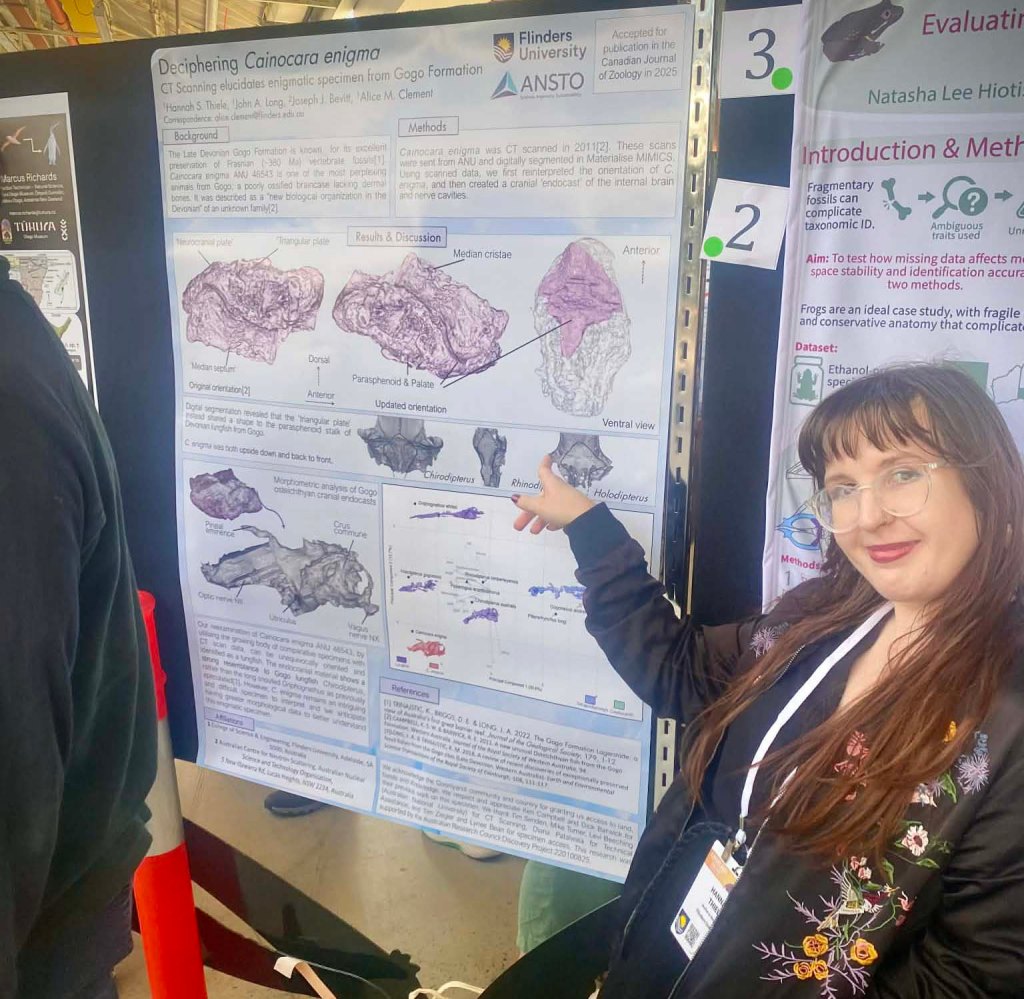
We deduced that the enigma from Gogo was most likely the poorly-preserved, and heavily-weathered braincase of a lungfish. Exactly which one we don’t know, but something similar to the short snouted forms such as Chirodipterus or Holodipterus.
Lead author, Hannah Thiele, presenting her results on the Gogo enigma at the CAVEPS meeting in 2025.
If you are keen to read more, see the news article “Missing pieces added to ancient global fish puzzle” published on Scimex. After this work, Hannah continued her study on fish brains at Flinders University, and has now commenced her PhD.
Nope, I am not about to talk about figures from the Hebrew Bible (you’re certainly in the wrong place if that is what you’re after), but instead some fantastic student-led work that was published recently by two of my students.
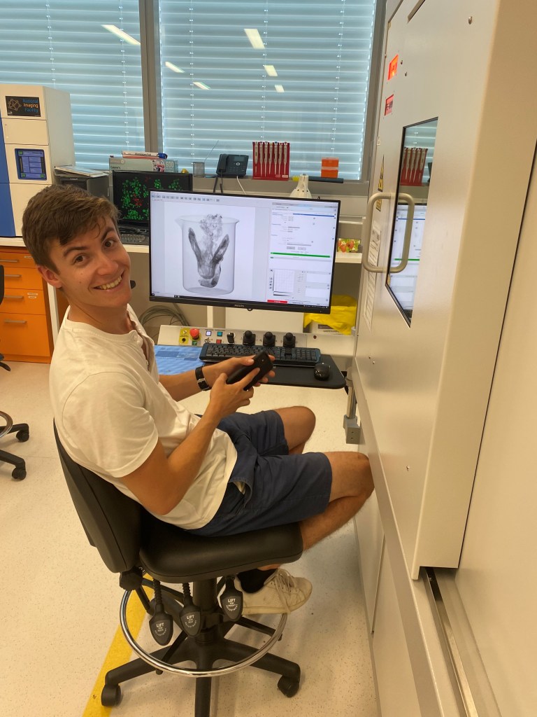
Joshua Batt posing with a fossil rhizodont.Joshua Bland CT scanning some fossil lungfish.
He worked on fossil lungfishes from the Late Devonian Gogo Formation, of northern Western Australia, which are beautifully preserved in 3D. Gogo is an interesting site for lungfishes, as this ancient tropical reef preserves the most diverse assemblage of lungfish species in any space or time from throughout their 400 million year history.
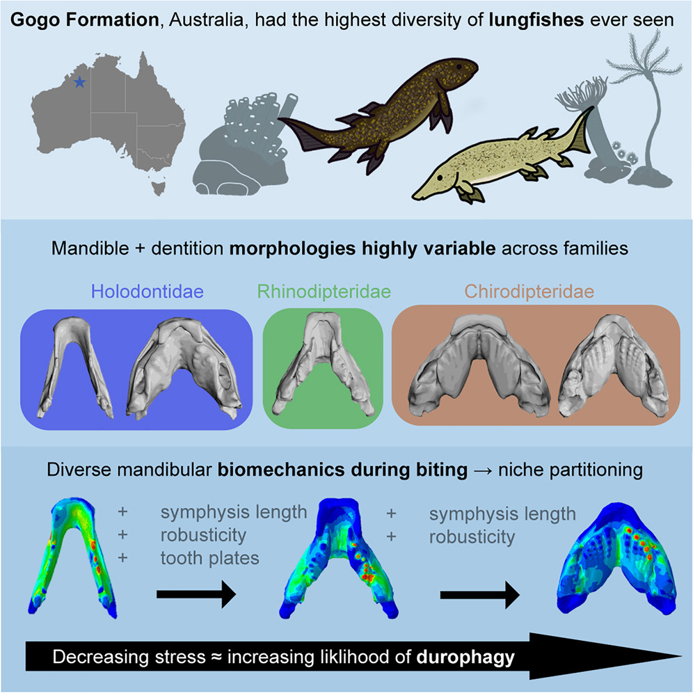
The lungfish at Gogo are taxonomically diverse (many species), but also morphologically disparate (anatomically different from each other). We wondered how this may impact function, did their jaws work biomechanically differently from one another?
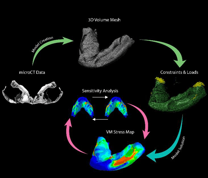
To answer this question, Josh used a technique adapted from engineering, called Finite Element Analysis (FEA). FEA is a computer-based method used to predict how structures respond to external forces by calculating the stress and strain within small, divided elements of the object. The first use of FEA in palaeontology was by Emily Rayfield and colleagues on the skull of a large theropod dinosaur in 2001, so its use remains quite recent for the field, and our study is the most comprehensive application to fossil fishes published thus far (yay!).
Generalised methods figure from Bland et al. (2025) showing stepwise process to solve Finite Element Models of fossil lungfish mandibles (lower jaws) during biting.
Together with our co-authors, Hugo Dutel, John A. Long, Matteo Fabbri, Joseph Bevitt, and Kate Trinajstic, and our very own FEA guru, Olga Panagiotopoulou, we found a diversity of stress and strain experienced by our lower jaw models, with some surprising results. This diversity of jaw morphology and biomechanics seen among the lungfish at Gogo may have been one of the reasons driving their great success. Different lungfish were adapted for eating very different things and so weren’t in resource competition with each other.
Our comprehensive dataset offers the most detailed quantification of biting performance in any fossil fish thus far, providing biomechanical evidence for diverse feeding adaptations and niche partitioning within Gogo lungfishes.
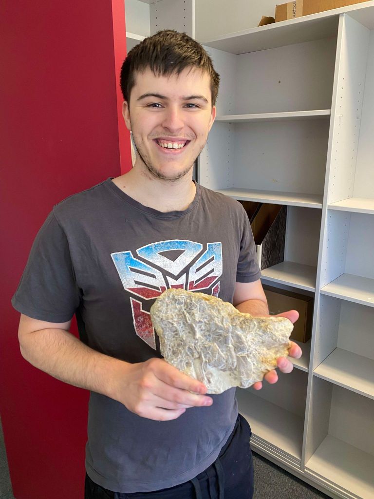
Working together with Dr Tom Challands, from the University of Edinburgh, researchers at Flinders University have been busy preparing and describing a real beast of a fish from the Early Carboniferous of the UK. The specimen was extracted by Tom and colleagues from Burnmouth in Scotland, but 10 or so large blocks of material came to Australia in 2020 for further study. Painstaking mechanical preparation has been done by Carey Burke and revealed what is likely to be one of, if not the most, complete articulated rhizodont fish known.
John Long, Carey Burke and Josh Batt inspect a specimen of fossil rhizodont at Flinders University.
Rhizodonts were large predatory lobe-finned fishes that lived throughout the Devonian and Carboniferous periods, usually in freshwater or estuarine environments. Some are thought to have grown as big as seven meters in length and they had very big teeth. Not a fish I would like to swim with!
Josh started working on this material for his Honours project during 2024, and there still remains a lot of it to be formally described, especially post-cranial material, but this first “rapid communication” paper presents the “main skull block”. Is this thing more beauty or beast… (perhaps beauty really is in the eye of the beholder?)
Image from Batt et al. (2025) JVP showing the dorsal view of NMS G.2025.10.1.1.
Needless to say, I felt very proud seeing two of my students have their first peer-reviewed publications come out. I expect more good things to come from both of them.
Want more? You can find both of these fantastic papers via the links below:
It seems I had such a “SPring” in my step, I completely forgot to write about my trip to Japan in November last year!
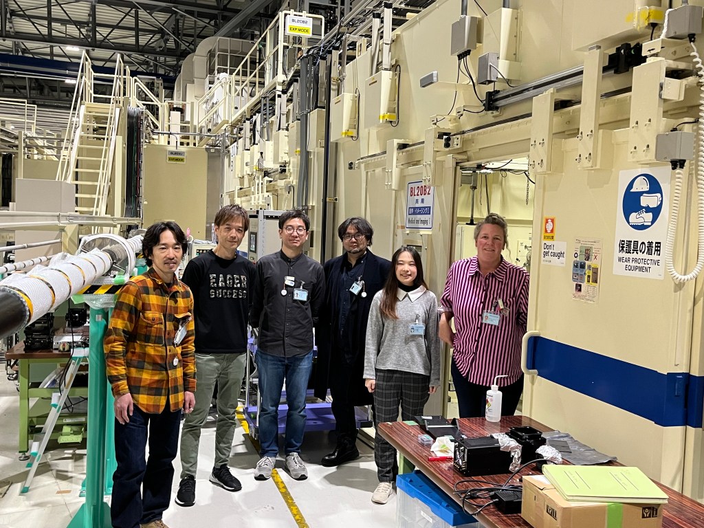
Thanks to a Flinders University International Research Engagement grant, I had the means to visit SPring-8 (the world’s largest third-generation synchrotron radiation facility) to work with my collaborator A/Prof. Tatsuya Hirasawa scanning embryos of the Australian lungfish, Neoceratodus forsteri.
The Japanese synchrotron, known as SPring-8, is nestled atop a mountain in Hyōgo Prefecture in the Kansai region of Japan. The nearest major cities are Osaka and Kobe. Sika deer amble through the grounds which are frequently shrouded in fog (they inspired me to write a haiku), and somewhat unusually for synchrotron facilities, there are paths to ride bicycles inside!
The team at SPring-8, Japan, after a successful experiment.
Sika deer through fog
SPring-8 autumnal mountain
Embryos revealed.
I was there working with Tatsuya Hirasawa and his team to image an ontogenetic (referring to the development of an organism throughout its lifespan) sequence of the Australian lungfish. I’ve previously worked on this animal to describe aspects of its brain (Clement et al. 2015) and muscle (Ziermann et al. 2017) anatomy (but see also Challands et al. 2020 where we discuss brains AND muscles in the same paper!)
Even though I had worked at the ANSTO Australian Synchrotron and ESRF in Grenoble before, I had never scanned tiny embryos of animals, and so learnt a lot about the preparation and parameters best suited to this kind of material. Other researchers working with us included Hiroki Higashiyama, as well as Toru Kawanishi and Kiiri Hama, respectively scanning either chicken embryos or bichir fish fins.
Left to right: Tatsuya Hirasawa, Masato Hoshino (beamline scientis), Toru Kawanishi, Hiroki Higashiyama, Kiiri Hama, and Alice Clement, using BL20B2 beamline at SPring-8, Japan.
Following on from our successful synchrotron experiment, I spent a week in Tokyo. I was honoured to give an Evolutionary Morphology seminar at the beautiful and historic campus of the University of Tokyo (those ginkgo leaves!) and visit Tatsuya Hirasawa’s lab. Tatsuya Hirasawa and his group analyse fossil specimens using synchrotron radiation, as well as developmental genetic analyses of living animals at the gene and cellular level to investigate “Evo-Devo” of vertebrates. To his group and other guests from various institutions in Tokyo, I presented research on “Digital Palaeontology of the Early Vertebrates” to a very engaged and interesting group.
Evolutionary Morphology seminar, University of Tokyo, Japan, November 2024.Alice and Tatsuya at the University of Tokyo.
I also took the opportunity to travel nearby Tsukuba to visit A/Prof. Daichi Suzuki (University of Tsukuba), whom I had met recently at the ISELV meeting in Quebec. He investigates the evolutionary origin of the vertebrate brain and consciousness, which is absolutely fascinating! Whilst there I got to look at some cool lamprey scans he and his students are working on, and give a palaeontology seminar. Biiiiiiiiiiiiiig thanks to the amazing Chisako Sakata for the tour of the National Museum of Nature and Science Tsukuba Research Center!
Visiting the National Museum of Nature and Science Tsukuba Research Center with my family.
Paleomorphology Seminar, University of Tsukuba.
I had such a wonderful time in Japan, AND was lucky enough to get back briefly less than two months after this visit for the 4th International Coelacanth Symposium, also held in Tokyo. Two visits in close succession was a great reason to revive my old high school Japanese… (また日本に行きたい!) That being said, I can’t wait for the next trip!!!