PalAss, Bristol

The final leg of my Europe trip took me to Bristol, in Somerset, England. The Annual Meeting for the Palaeontological Association was held in and around the University of Bristol and the city Museum over four days this December.

The conference kicked off with a special symposium “Frontiers and Advances in Dinosaur Palaeobiology” on Friday in the impressive Great Hall in the Wills Memorial building, followed by regular sessions over the weekend. The social programme was almost as busy as the scientific one, with many opportunities to socialise and network with fellow palaeontologists.

PalAss

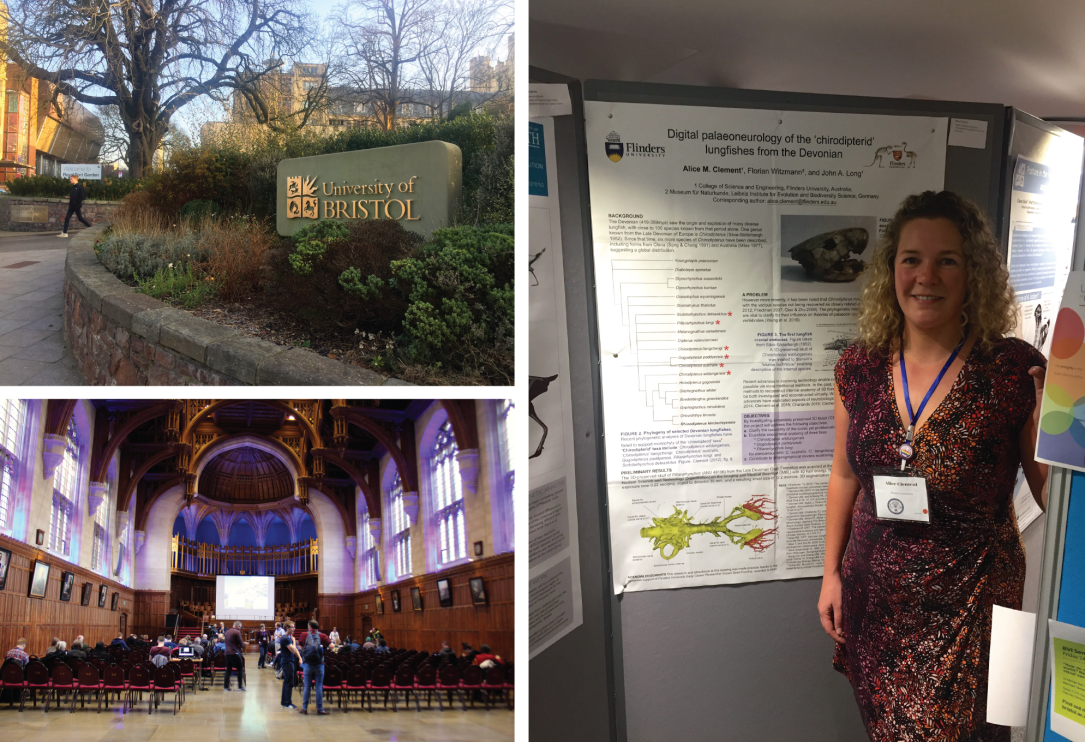
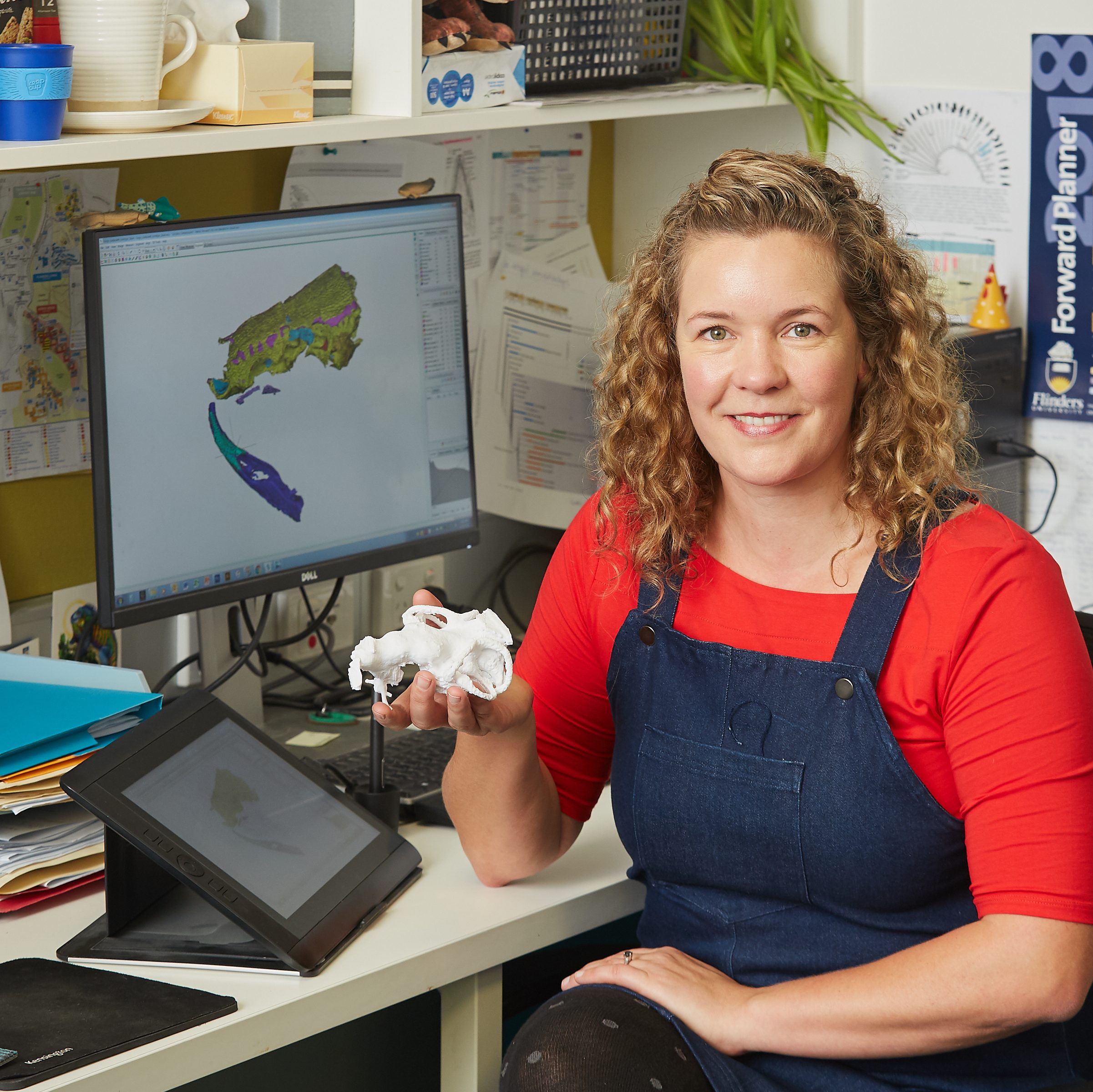
I presented a poster on the “Digital palaeoneurology of the ‘chirodipterid’ lungfishes from the Devonian” – outlining one of my current projects on the fossil brains of an iconic group of fossil lungfish. It was my first experience presenting a poster rather than an oral presentation, and as such, I found the whole experience much more relaxing than usual!

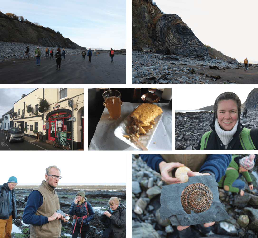
Monday took us to the Somerset coast on a cold but clear day to inspect the Triassic-Jurassic section on the beach at Watchet. We searched for beautiful nacre “mother-of-pearl” ammonites and ichthyosaur bits, then enjoyed traditional fish and chips at the local pub and a pint (or two) of delicious cider. Many thanks to Dr Jakob Vinther (bottom left image) and colleagues for a very well organised, highly informative and fun meeting and field-trip.

Fieldtrip

ESRF, Grenoble

What would you like to do on your 30th birthday? Shut down for two years and replace your major organs? It may not sound like an ideal celebration, but that is what the European Synchrotron Radiation Facility (ESRF) will do this December.

The world’s first third-generation synchrotron light source (ESRF) was built in 1988, and will pause the world’s “most intense X-rays for research” to enable the construction of a new storage ring and the addition of even more beam lines. The Extremely Brilliant Source (EBS) is not scheduled to come back online until late 2020.

I feel especially lucky to have been here in beautiful Grenoble this week working with colleagues from Uppsala University, Sophie Sanchez and Per Ahlberg, on the very final experiment on beamline ID 19 before the scheduled shutdown. ESRF is the only synchrotron in the world with a beam powerful enough to perform this experiment at the super high resolution required (down to 0.7μm voxel size!).

IMG_4637Per and Sophie inspecting a mounted specimen on the ID19 beamline at ESRF

During 72 hours of beam time, we work 24/7 with help from ESRF researcher, Paul Tafforeau, to scan as many specimens as possible. Sophie’s project is looking at the bone histology of the fins and limbs over a number of significant evolutionary transitions: the fin-limb, water-land, and the characteristics that appear in the first amniotes (animals that lay a waterproof egg, e.g. the first reptiles).

Bone histology, microanatomy and skeletochonology can be remarkably informative about the lifestyle and life history traits of an animal. For example, the bone microstructure can indicate whether an animal matured quickly, or had a long juvenile phase – as shown recently by Sophie and colleagues in Sanchez et al. (2016) and other works.

Untitled-1Alice setting up a specimen (left), and Laugia, a Triassic coelacanth from Greenland (right), scanned at the ESRF

Skeletochronology works on the same concept as counting tree rings in trees, and can map various changes in life history, development and physiology inside the bones of an animal. And very significantly for these experiments, the structure of the bone can indicate whether the bone was capable of sustaining an animal’s weight on land, or if it must have remained buoyant in the water.

It’s been an exhausting yet exciting few days scanning many stunning specimens sourced from all over the world. I look forward to continuing our work on these projects in the coming years (and catching up on some sleep!)

IMG_4617
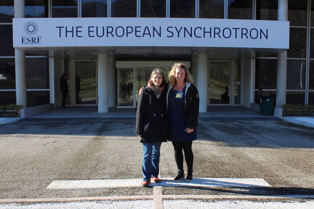
Alice and Sophie at the ESRF in December 2018

Berlin Museum für Naturkunde

I’ve had a wonderful few days working at the Berlin Natural History Museum. I was interested to see the original specimen of a lungfish (Chirodipterus wildungensis) that was the first to have the internal space for the brain described. This was achieved over 65 years ago using the “shatter method” (which is destructive as it sounds!)

Berlin

I am also interested in a small coelacanth skull, called Euporosteus eifeliensis, from the Devonian of Germany (~383-388 million years ago). It is preserved in 3D and is only known from a single specimen!

To study both the fossil lungfish and coelacanth, I used the traditional method of making observations using a microscope, but also will 3D model them both using the museum in-house CT scanner and segmenting software back in Adelaide.

Florian

I’m so very grateful to Dr. Florian Witzmann, curator of fossil fish and amphibians, and Dr. Kristin Mahlow, who works in the CT lab, for their assistance this week. I’m looking forward to continuing our collaboration together.

Another perk of working in the museum was the opportunity to browse through their galleries, which include a beautiful Archaeopteryx specimen on display, as well as a visiting T-rex skeleton called Tristan. As one of the best preserved and most complete T-rex skeletons in the world, he was very impressive!

Trex