New paper out today!

Today, my latest paper investigating a 400 million year old fish, has been published in the journal eLife. Together with my colleagues from Australia, the UK, Sweden, and the Netherlands, we have studied and uncovered new information about an enigmatic fish known as ‘Ligulalelpis.

There are just two specimens of this fish’s skull known, and we have examined both of them using microCT to view the internal anatomy in addition to the external features.  By doing so, we have settled a 20 year controversy surrounding this animal, and identified it as belonging just below the major radiation of all modern fish, amphibians, reptiles, birds and mammals (that’s 98% of all vertebrate species alive today!) on the evolutionary family tree.

In addition to the eLife article, you can read a piece written by John Long and myself for The Conversation, including some lovely images from Brian Choo.

Ligulalepis

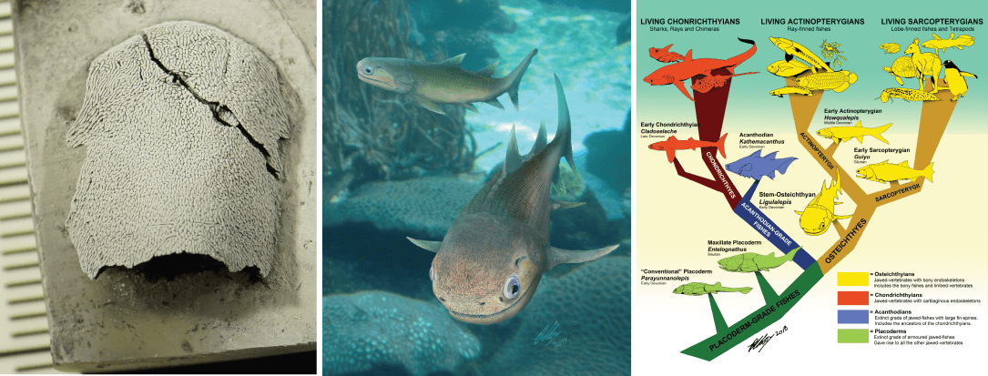
Figure. A, the new skull viewed from above (dorsal view); B, life reconstruction of ‘Ligulalepis‘; and C, the position of ‘Ligulalepis‘ in the evolutionary family tree.         (Photo and animation below, Ben King; Illustrations, Brian Choo.)

***ALSO: below is a short video with John and myself, talking about ‘Ligulalepis‘, filmed by Yaz Dedovic at Flinders University Paleontology Labs, Adelaide.

DiceCT

My diceCT work on the Australian lungfish has been featured at the online home of the diceCT community. DiceCT stands for:

Diffusible

Iodine-based

Contrast-Enhanced

Computed

Tomography,

and is a relatively recent technique for imaging soft tissue using CT imaging, and it gives spectacular results!

The site is a great place to stay up-to-date on recent publications using the method, technical advice and events. You can also read about other techniques, such as the deliciously-named ‘SpiceCT’ (Selectively Perfusable Iodine-based Contrast-Enhanced CT), which is particularly good for staining large specimens, and can do so very rapidly.

See the original blog post and other cool work using these methods via New Publication: Cephalic muscle development in the Australian lungfish, Neoceratodus forsteri.

My diceCT publications on the Australian lungfish:

  • CLEMENT, A. M., NYSJÖ, J., STRAND, R. & AHLBERG, P. E. 2015. Brain – endocast relationship in the Australian lungfish, Neoceratodus forsteri, elucidated from tomographic data (Sarcopterygii: Dipnoi). PloS One.
  • ZIERMANN, J. M., CLEMENT, A. M., ERICSSON, R. & OLSSON, L. 2017. Cephalic Muscle Development in the Australian Lungfish, Neoceratodus forsteri. Journal of Morphology, 279, 494-516.

Latimeria love!

I’m in the eastern cape of South Africa at the moment, working with Rhodes University palaeontologist, Dr Rob Gess. Rob has been working on a site called Waterloo Farm which has yielded many spectacular fossil discoveries … but more on this later.

  • Read about the new species of fossil lungfish that Alice and Rob described from Waterloo Farm HERE.

Fans of the coelacanth Latimeria, would know this part of the world well. In 1938, fishermen hauled up a strange fish from the oceanic depths off the coast of South Africa. This fish was identified as a coelacanth, and given it’s name in honour of Marjorie Courtenay-Latimer, curator of the East London museum who helped to identify the fish.

Coelacanth’s belong to the Sarcopterygii (“lobe-finned” fish), the group that also includes lungfish (wooo!) and tetrapods (the first land-living vertebrates and all their descendants – including you). Today they live at great depths, have a strange hinge separating the front and back parts of their skulls, give birth to live “pups”, and have a special electroreceptive organ in their snout.

Previous to the 1938 discovery, the coelacanth lineage was thought to have gone extinct some 70 million years prior, around the same time of the dinosaurs. Latimeria caused quite a stir upon discovery – it was nicknamed “Old Four Legs” and people thought it was the direct ancestor of mankind! (Spoiler: it is not). Unfortunately the first specimen identified was rotten inside before scientists could dissect it. So the hunt was on for a second specimen… which took a long 14 years!

It is this second specimen, discovered in the Comoros in 1952 and the first one to be dissected, that is on display at the South African Institute for Aquatic Biodiversity here in Grahamstown, South Africa (formerly named the JLB Smith Institute of Ichthyology, after the man who named and described Latimeria). I went to pay my respects to this mighty lobe-finned fish yesterday. Latimeria loooooove!

IMG_3191