This week I visited the newly-built and impressive-looking SAHMRI (South Australian Health and Medical Research Institute) building in Adelaide.
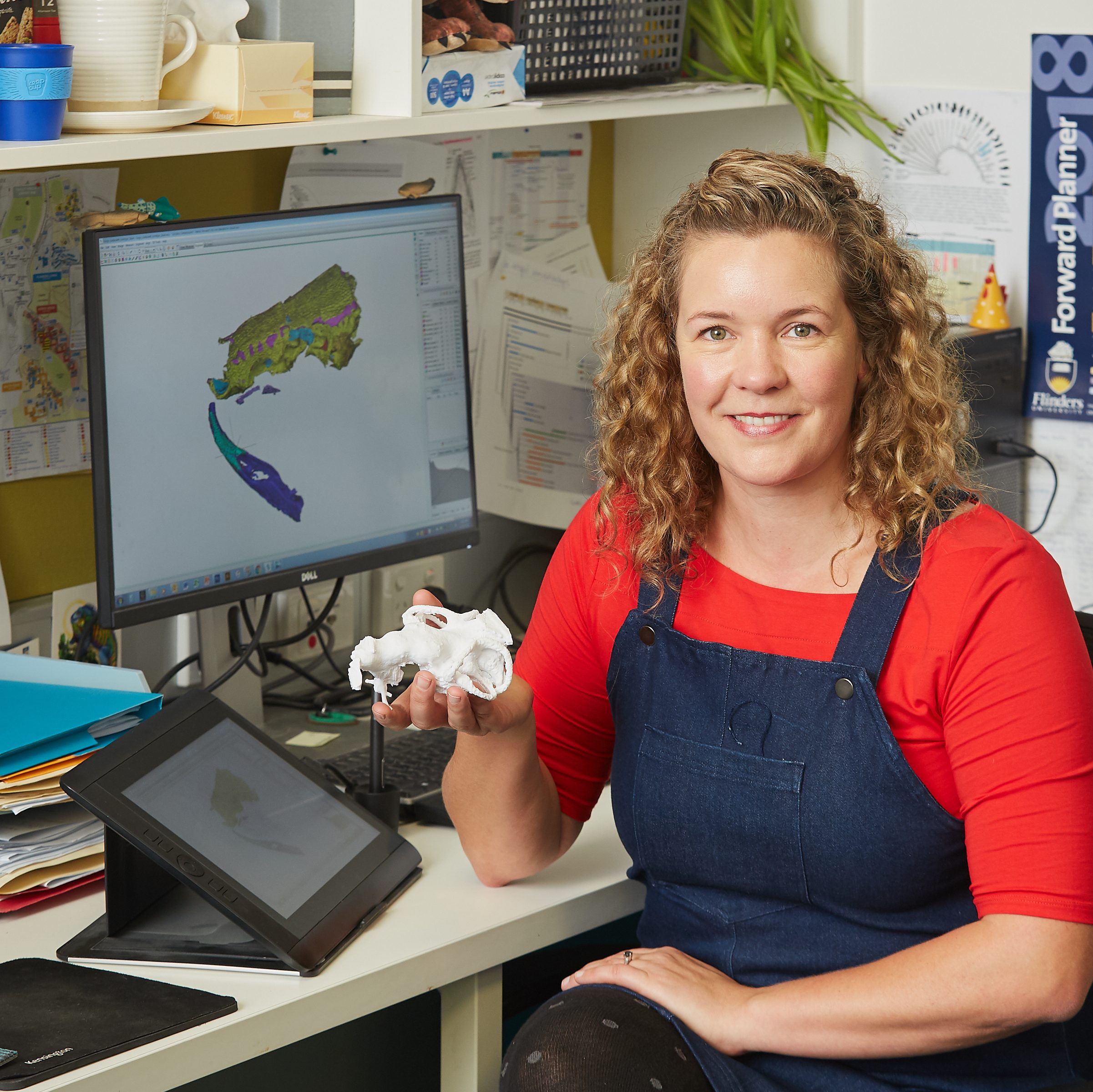
Together with Trevor Worthy, Warren Handley and Phoebe McInerney, we used their hospital CT scanner to scan the tracheal system of a cassowary. This is in aid of Phoebe’s Honours project, which I am co-supervising.
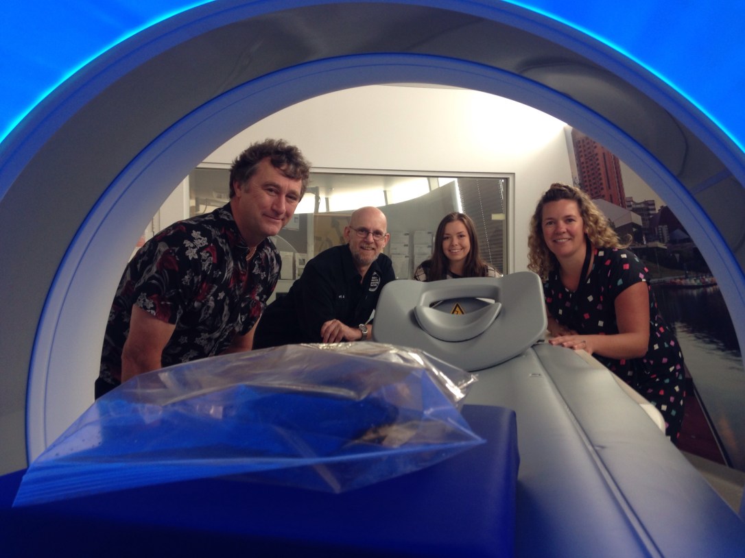
Figure 1: Trevor, Warren, Phoebe and I (and cassowary) at SAHMRI this week.
We are using a technique called diceCT which stands for “Diffusible Iodine-based Contrast Enhanced Computed Tomography”. It is a relatively new technique that yields spectacular results (see Figure 2). It is mostly being used by morphologists who want better differentiation between different types of soft tissues than you normally get from a regular CT scan – the results are comparable with those you get using MRI (Magnetic Resonance Imaging).
A big thank you to Mishelle! We were very excited to visit the new facility, and I look forward to seeing Phoebe’s project take shape throughout the year.
- Update: read the publication by McInerney et al. (2019) HERE
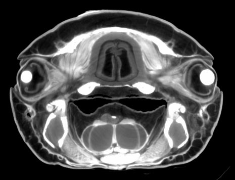
Figure 2: The head of the Australian lungfish, Neoceratodus, imaged using diceCT.

1 thought on “DiceCT & Scanning at SAHMRI”